|
Lääketieteellisen fysiikan ja tekniikan yhdistys (LFTY) Finnish Society for Medical Physics and Medical Engineering |

IX Finnish Medical Physics and Medical Engineering Day, 10.2.2010, Tampere University of Technology
The ninth Finnish Medical Physics and Medical Engineering Day was held 10 February 2010 at Tampere University of Technology, Tampere, Finland. The annual event gathered this year 88 student and researcher participants and five Finnish companies from the area of medical physics and medical engineering. The traditional poster exhibition, where the best Master's theses and diploma theses finished in 2009 were awarded had 14 participants. The award for the best theses was this year 2009 &euro.From the 14 participants, two theses were reckoned to be above the others. These two theses were:
- Aliina Tuomenlehti, Tampere University of Technology
EEG interface for portable ECG recorders - Panu Vesanen, Aalto University School of Science and Technology
Compressed Sensing in Parallel Magnetic Resonance Imaging
- Tuomas Koivumäki, Tampere University of Technology
Theoretical background for respiratory and cardiac gating in SPECT/CT using electrical bioimpedance method - Markus Malo, Tampere University of Technology
Numerical analysis of uncertainties in dual frequency ultrasound technique - Tuomas Viren, University of Eastern Finland
Nivelensisäinen ultraäänitekniikka nivelrikon diagnostiikkaan
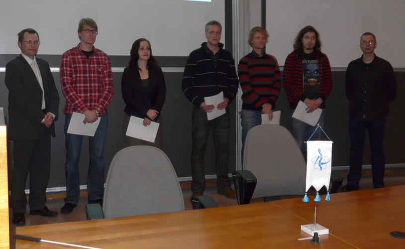 |
| Award presentation for the best Master's theses and diploma work in 2009 (from left to right): Prof. Pasi Karjalainen (chairman of Finnish Medical Physics and Medical Engineering Society), Tuomas Koivumäki, Aliina Tuomenlehti, Markus Malo, Panu Vesanen, Tuomas Viren ja Mika Tarvainen (secretary of Finnish Medical Physics and Medical Engineering Society).. |
Abstracts of the awarded theses
EEG interface for portable ECG recorders
Aliina Tuomenlehti, Tampere University of Technology
A number of different medical conditions can cause unconsciousness. Epilepsy and trauma are examples of such conditions. These disorders affect the normal electrical brain activity and in clinical medicine these conditions are often diagnosed using electroencephalogram (EEG). Because of the complexity of the measurement procedure, EEG is not generally used in emergency medicine. However by utilizing EEG in emergency situation, vital information could be obtained.
The purpose of this thesis was to develop an interface enabling EEG measurement in emergency medicine. In order to make the implementation as simple as possible, the interface was designed to be compatible with a monitor used in electrocardiogram (ECG). Because the characteristics of the EEG and the ECG signals differ, the designed interface should be able to adapt the EEG signal for the monitor. This constitutes the basis for the system specifications. Because the EEG signal is small-amplitude, the interface should provide adequate gain. The noise level should be kept as low as possible, because such unwanted signal can interfere or even overrun the desired signal. Small size was one of the main objectives in this project, because the size has a great effect on the usability of the system. In order to make the device portable, battery was used as a power supply. This feature and the desired 24 hour operation time, required low power consumption.
The interface is based on a microcontroller unit (MCU) thus the system can be referred as an embedded system. This thesis covers the hardware design while the software development is done separately. The system is divided into operational blocks and the prototype development for clinical evaluation testing is done based on these. The hardware design was done based on both the initial system specifications and the aim of continuing improvement. Operational tests were done with an actual ECG-monitor to ensure the comparability with the actual measurement. Physiological signals like ECG and EEG were used in order to verify the operation of the interface.
All of the before mentioned specifications were fulfilled. Test measurements made during this project showed that the developed interface acts as an adapter which compensates the differences between the EEG and ECG signals so that the usage of the ECG-monitor is possible. Tests also showed that by using the designed interface good-quality EEG signal can be measured. From this signal it is possible to distinguish variations which indicate disorders affecting the brain activity and causing unconsciousness. It is evident that this would improve the quality of the emergency care.
Compressed Sensing in Parallel Magnetic Resonance Imaging
Panu Vesanen, Aalto University School of Science and Technology
Introduction and objective of the work
Magnetic resonance imaging (MRI) is a non-invasive method that allows the study of interior structures of matter. MRI is based on magnetizing a sample, manipulating the magnetization, and detecting the magnetic field that the sample produces. Today, MRI is widely used in medical imaging. The goal of this work is to reduce MRI scanning times. Reductions in the measurement time are important because they correspond to, e.g., higher throughput of MRI scanners or enhanced spatial resolution of images.
Methods
It is well-known that natural images, such as MR images, are compressible. Compressed sensing (CS) is a method that exploits the compressibility of signals in order to measure and reconstruct them efficiently. Moreover, in parallel MRI (pMRI), the magnetic fields produced by the magnetized sample are measured with multiple coils simultaneously. The differing sensitivity profiles of the coils allow a faster rate of information flow compared to single-coil receiving. In this work, CS and pMRI are combined to achieve shortened measurement times.
Results
Simulations were conducted to demonstrate the performance of the method in idealistic conditions. Several different sampling patterns were attempted in the simulation. In addition, experimental measurements were performed to validate the technique in practice. Simulation results indicate that the combination of CS with pMRI reduces the measurement time by 3050%; truly randomized sampling patterns provide a slight acceleration as well compared to Cartesian ones. Experimentally, scanning time reductions of 1020% were obtained.
Conclusion
In this work, it is shown that the combination of CS with pMRI is feasible. Both simulation and experimental results indicate that the scanning time reduction using this technique is significant. Future work includes more advanced pulse sequences for CS implementation in pMRI as well as new, compressible, and MRI-compatible function bases for more efficient CS.
Theoretical background for respiratory and cardiac gating in SPECT/CT using electrical bioimpedance method
Tuomas Koivumäki, Tampere University of Technology
Motion due to respiration and cardiac function often produces artefacts on SPECT/CT images. Motion artefacts may lead to false diagnosis, for example, in evaluation of coronary artery disease. The effects of motion on image quality can be minimized by gating that is, the synchronization of image acquisition to a physiological function.
Respiration and cardiac function can be monitored utilising bioimpedance measurement methods. Impedance pneumography and cardiography are generally regarded as cheap, safe and reliable. The aim of the thesis was to establish foundation for bioimpedance-based simultaneous respiratory and cardiac dual-gating in SPECT/CT. The main objective was to determine an optimized electrode configuration for simultaneous measurement of respiration and cardiac function using one measurement configuration that is, a pair of current injection electrodes and a pair of voltage measurement electrodes. A further objective was to study the requirements of signal processing for the separation of both gating signals from a single bioimpedance measurement signal.
In order to determine the most sensitive electrode configuration for simultaneous detection of respiration and cardiac function, computerized finite element method (FEM) modelling was performed. FEM calculations were carried out using a simplified human upper thorax model with CT-based inhomogeneities (heart, lungs and spine) and realistic tissue conductivities. The performance of over 47 000 electrode configurations was computed utilising the complete electrode model of electrical impedance tomography (EIT). The results were analyzed in terms of sensitivity and selectivity for the anatomical regions of heart and lungs. In order to study bioimpedance measurement dynamics, forward problem was solved to simulate measurements with feasible electrode configurations. Respiration and cardiac function were imitated by the alteration of lung and heart conductivities, respectively. Finally, the separation of respiratory and cardiac gating signals from simulated measurement data was studied.
An optimized electrode configuration for simultaneous detection of respiration and cardiac function was located on the level of fourth and fifth intercostals spaces on the anterior thorax. One electrode of both electrode types is located in midway and 1/3 of the distance from sternum to axilla on left and right sides, respectively. The absolute values of sensitivity for the determined electrode configuration were 75 % and 69 % of the sensitivities of separately optimized electrode configurations for respiration and cardiac function, respectively. The forward problem simulations and signal separation analysis indicated that signals for cardiac and respiratory gating can be obtained performing only one bioimpedance measurement.
The results of this study clearly indicate that the bioimpedance method is potential for dual-gating of SPECT/CT imaging. Since only four electrodes are needed to implement the method, it is simple to apply clinically and comfortable for the patient.
The thesis study was the first phase of a research project, which strives to develop a clinically feasible bioimpedance-based dual-gating method and equipment for nuclear medicine imaging.
Numerical analysis of uncertainties in dual frequency ultrasound technique
Markus Malo, Tampere University of Technology
Quantitative ultrasound (QUS) measurements have shown potential in the diagnostics of osteoporosis. However, the variation in the thickness and composition of the overlying soft tissues results in significant errors in the bone QUS parameters and diminishes the reliability of the technique in vivo. Recently, the dual frequency ultrasound (DFUS) technique was introduced to minimize the soft tissue induced errors.
In this study, the significance of soft tissue induced errors and their elimination with the DFUS technique were simulated using a finite difference time domain technique. Furthermore, we investigated the potential of the DFUS corrected integrated reflection coefficient (IRC) of bone to detect changes in the cortical bone density. The effects of alterations in the thickness of fat and lean tissue layers and the inclination of interfaces between the soft-tissues and between the soft tissue-bone layers were simulated. In addition, the errors arising from non-optimal focusing of the transducer to the soft tissue-bone interface were investigated.
The main findings of this masters thesis were that when the angle of the soft tissue interface was zero, i.e. perpendicular to the incident ultrasound beam, the DFUS-calculated soft tissue composition correlated highly linearly with the true soft tissue composition. The inclination between the soft tissue-bone interface was found to be critical. Even a 2-degree inclination between the soft tissue and the bone surface induced an almost 18% relative error in the corrected IRC. Increasing the inclination between the soft tissue layers increased the error in the DFUS-calculated lean and fat tissue thickness. This error was especially significant at inclination angles greater than 20 degrees. The significant soft tissue induced errors in IRC values (> 300 %) could be effectively minimized (< 10 %) by means of the DFUS correction. Importantly, after the DFUS correction, physiologically relevant variation in the cortical bone density could be detected (p < 0.05). In the out-of-focus study, the average relative error in IRC values was more accurate after the attenuation correction was done with the DFUS-calculated soft tissue thicknesses, than with the actual soft tissue thickness from the simulation geometry (8.9 % vs. 28.7%).
The study showed that with the DFUS technique one can accurately determine the thickness of the soft tissues overlying the bone surface, provided that each tissue layer interface is perpendicular to the incident ultrasound wavefront and the soft tissue-bone surface is in focus. This enabled us to correct accurately the reflection arising from the soft tissue-bone interface. Even small physiologically relevant differences in the cortical bone density could be separated with DFUS-corrected IRC values. The study showed that the more the interfaces of the tissues are inclined, the greater is the error induced in the DFUS-calculated thicknesses as well as on the attenuation correction of the reflection from the soft-tissue-bone interface. The changing thickness of the soft tissue layer, which leads to non-optimal focusing, was responsible for introducing an error in the DFUS calculations, when each tissue layer interface was perpendicular to proceeding ultrasound wavefront and a fixed focal length transducer was used.
Nivelensisäinen ultraäänitekniikka nivelrikon diagnostiikkaan
Tuomas Viren, University of Eastern Finland
Nivelrikko on yksi yleisimmistä syistä nivelkipuun ja liikuntakyvyn alenemiseen. Nivelrikon tyypillisiä oireita ovat nivelrustopinnan rappeutuminen sekä rustonalaisen luun paksuuntuminen ja epämuodostumat. Nivelrikkoa ei vielä voida parantaa ja nykyään sen hoito perustuu kivun lievittämiseen. Kuitenkin, jos nivelrikko havaitaan riittävän aikaisessa vaiheessa, voidaan sen etenemistä mahdollisesti hidastaa elämäntapamuutoksilla sekä lääkehoidolla. Nykyisillä kliinisessä käytössä olevilla tekniikoilla nivelrikko voidaan havaita vasta, kun rusto on peruuttamattomasti vaurioitunut. Kirjallisuudessa on esitetty useita kokeellisia, ei vielä kliinisessä käytössä olevia, tekniikoita joilla pystytään havaitsemaan ensimmäisiä nivelrikolle tyypillisiä kudosmuutoksia. Kvantitatiivinen ultraäänikuvantaminen on yksi lupaavimmista uusista tekniikoista.
Tässä opinnäytetyössä jatkokehitetään korkeataajuista ultraäänimenetelmää nivelrikon diagnostikkaan soveltamalla ensimmäistä kertaa kliinisessä käytössä olevaa IVUS-ultraäänilaitetta nivelruston kvantitatiiviseen kuvantamiseen. Työn tavoitteina oli tutkia, voidaanko IVUS-laitteella havaita eroja keinotekoisten testikohteiden pinnankarheudessa sekä tutkia voidaanko menetelmällä erottaa mekaanisesti tai biokemiallisesti vaurioitetut rustonäytteet koskemattomista näytteistä. Tutkia menetelmän soveltuvuutta nivelensisäiseen ultraäänitutkimukseen tekemällä naudan polven niveltähystys laboratorio-olosuhteissa.
Työssä tutkittiin minimaalisesti invasiivisen ultraäänitekniikan soveltuvuutta nivelrustopinnan vaurioiden havaitsemiseen laboratorio-olosuhteissa. Terveitä (n = 13), mekaanisesti vaurioitettuja (n = 7) sekä entsymaattisesti vaurioitettuja (n = 6) naudan rustonäytteitä ja erilaisia keinotekoisia testikohteita kuvattiin korkeataajuisella (40 MHz) ultraäänilaitteella. Kaikille mitatuille rustonäytteille sekä testikohteille laskettiin ultraäänen heijastuskerroin (Reflection coefficient, R), integroitu heijastuskerroin (Integrated reflection coeffisient, IRC) sekä pinnankarheusindeksi (Ultrasound roughness index, URI). Lisäksi tutkittaessa tekniikan kliinistä soveltuvuutta yksi terve naudan polvinivel kuvattiin niveltähystystä jäljittelevissä olosuhteissa.
Testikohteiden pinnankarheuksien erot havaittiin kaikilla ultraääniparametreilla. Rustopinnasta mitatut heijastuskertoimet pienenivät merkittävästi (p < 0.05) sekä mekaanisen että entsymaattisen vaurioituksen jälkeen. Lisäksi rustopinnan karheus suureni merkittävästi (p < 0.05) mekaanisen vaurioituksen jälkeen.
Tämä tutkimus osoitti että nivelensisäisellä ultraäänitekniikalla voidaan havaita sekä mekaanisesti että entsymaattisesti aiheutettu vaurio nivelrustossa. Lisäksi tekniikan soveltaminen kliinisen niveltähystyksen yhteydessä todettiin teknisesti mahdolliseksi.