|
Lääketieteellisen fysiikan ja tekniikan yhdistys (LFTY) Finnish Society for Medical Physics and Medical Engineering |

XI Finnish Medical Physics and Medical Engineering Day, 9.2.2012, University of Turku / Turku University Hospital
The 11th Finnish Medical Physics and Medical Engineering Day was held 9 February 2012 at Turku University Hospital, Turku, Finland. The annual event gathered this year 77 student and researcher participants. The traditional poster exhibition, where the best Master's theses and diploma theses finished in 2011 were awarded had 14 participants. The award for the best theses was this year 2011 €.From the 14 participants, one thesis was reckoned to be above the others. This thesis was:
- Andrei D. Jakab, Tampere University of Technology
Life-saving EEG system for use in emergencies
- Joanna Pylvänäinen, University of Turku
Transnedothelial migration of breast cancer cells during cancer metastasis - Koos C.J. Zevenhoven, Aalto University
Towards high-quality ultra-low-field MRI: novel methods for suppressing eddy currents
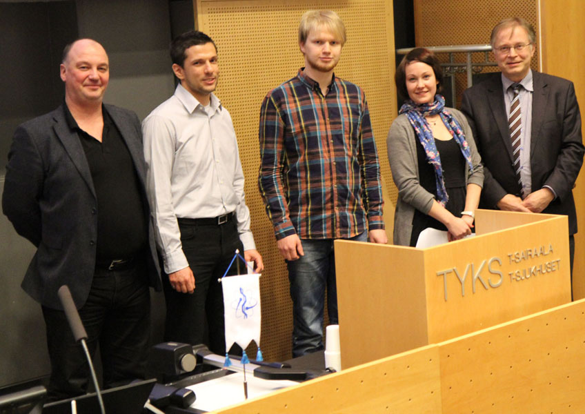 |
| Award presentation for the best Master's theses and diploma work in 2011 (from left to right): Prof. Jari Viik (vice-chair of Finnish Medical Physics and Medical Engineering Society), Andrei Jakab, Koos Zevenhoven, Joanna Pylvänäinen and Prof. Risto Ilmoniemi (chairman of Finnish Medical Physics and Medical Engineering Society). |
Abstracts of the awarded theses
Life-saving EEG system for use in emergencies
Andrei D. Jakab, Tampere University of Technology
Objective: The World Health Organization (WHO) classifies neurological disorders as a great threat to public health since these disorders and their sequelae are estimated to affect approximately one billion people worldwide. One of the most commonly used tests for the evaluation of neurologic disorders is the electroencephalogram (EEG). Despite its proven usefulness, because traditional EEG diagnostic methods are relatively time-consuming, require specialized personnel, and the recording systems are cumbersome, the EEG remains underused in clinical practice and particularly in emergency medicine (EM), where a quick diagnosis is sought after. The objectives of this study were to design and implement two EEG recording systems that would address the specific needs of EM by enabling caregivers to quickly secure a correct diagnosis and to establish a treatment plan in the vital early stages.
Methods: Two avenues of research were pursued. The first explored the suitability of a 6-channel wireless recording system that interfaces with a portable computer. The resulting system consists of a quick-application cap that can be normally applied on the patient in under 3 min, a wireless EEG (WEEG) amplifier, which digitizes the EEG and transmits it wirelessly to a measurement computer, and recording software, which stores the digital EEG in the standard EDF+ format. The second was centred on the novel concept of allowing the EEG to be recorded using the same infrastructure that is currently used for the recording of electrocardiographic (ECG) signals. This small EEG-to-ECG adapter dynamically processes the EEG signal in order to allow it to be displayed on any available ECG monitor.
Results: A thorough validation of the systems demonstrated the recording of high-quality signals as well as reliable operation. In addition to verifying their electrical characteristics, the EEG recorded by both systems was compared to that of commercial systems and found to be equivalent. Moreover, the WEEG system successfully underwent healthy volunteer testing in an examination room at the Department of Clinical Neurophysiology and in the ICU of the Seinäjoki Central Hospital.
Conclusions: Two fully-working EEG systems suitable for recording in emergency situations with a minimal preparation time have been developed, and their quality demonstrated. These novel systems facilitate the measurement process and make it possible to expand the practice of EEG to new recording locations. Clinical studies with the WEEG system currently underway at the Seinäjoki Central Hospital will help establish the utility of portable EEG systems in other clinical specialities (e.g., polysomnography).
Transnedothelial migration of breast cancer cells during cancer metastasis
Joanna Pylvänäinen, University of Turku
Transendothelial migration (TEM) is a well known process in which a cell penetrates the endothelium of a blood or a lymphatic vessel. The most studied cell type that utilizes this mechanism is the leukocytes, which follow the chemokine gradient created by local inflammations and travel through the bloodstream towards the inflamed area. Once they reach the inflammation site, leukocytes penetrate the endothelium using one of two distinct routes, the paracellular or the transcellular route. Invasive cancer cells have shown to use similar machinery to penetrate vascular endothelium during cancer metastasis. The blood system has been considered as the main cancer metastasis route, but evidence of metastasis through lymphatic system is increasing, though the mechanism still remains unknown. The endothelium has commonly been considered as a passive mechanical barrier during TEM. However, recently it has been shown to be an active member of the cell invasion machinery, conveying signals from the invasion site into the cell and remodeling the cytoskeleton, therefore enabling TEM.
In this thesis, the invasion routes of invasive and non-invasive breast cancer cells through lymphatic and vascular endothelia were studied. Also the reorganization of endothelial cytoskeleton during TEM was followed. The main methods used for this research were confocal microscopy combined with advanced cell staining techniques.
The results suggested that in lymphatic endothelium cancer cell invasion happens predominately through paracellular route, whereas in vascular endothelia the invasion mainly occurs transcellularly. The cytoskeleton seems to play an important role in guiding the invading cancer cell to appropriate invasion site and enabling the invasion.
In conclusion, both lymphatic and vascular endothelia act as a dynamic member of the transendothelial migration machinery, although distinct endothelium specific properties remain. These properties were adapted according to the invading cancer cell type.
Towards high-quality ultra-low-field MRI: novel methods for suppressing eddy currents
Koos C.J. Zevenhoven, Aalto University
Introduction and aim of study. While the state of the art of magnetic resonance imaging (MRI) has developed towards using multiple-tesla magnetic fields, another approach has emerged, where the signal is detected by highly sensitive SQUID sensors in a magnetic field on the order of 100 µT. Such ultra-low-field (ULF) MRI has many promising characteristics, including safety, silent operation, potentially low cost, and compatibility with other electromagnetically sensitive technology. However, ULF MRI has not yet reached its breakthrough, as the image signal-to-noise ratio (SNR) and field of view (FOV) are still insufficient for most applications. The aim of this thesis work was to improve the image quality of ULF MRI by designing and implementing upgrades to the pioneering ULF-MRI setup at UC Berkeley.
Methods. Since, in ULF MRI, the signal is proportional to a prepolarizing field Bp, which is pulsed using a designated coil, a natural approach was to increase Bp and the associated FOV by installing a larger water-cooled coil. These changes, however, directly lead also to larger unwanted transient effects, most notably, eddy currents in the conducting walls of the magnetically shielded room (MSR), which screens the system from external magnetic interference. Therefore, an MSR with weakly connected regions was designed and built to accommodate shielding currents but no harmful transients. A quantitative theory to describe eddy currents in MSR walls was presented. Based on the theory, a novel method, dynamic shielding, was introduced, where a single additional coil is pulsed with current waveforms specifically designed to kill selected spatiotemporal components of the harmful transient.
Results. The amplitude of the transient magnetic field generated by eddy currents was reduced by three orders of magnitude at roughly 15 ms after a Bp pulse, meeting the MSR design criteria. The transient eddy current patterns in the walls of the old and new MSR were also reconstructed as a function of time from a large number of magnetic-field measurements. The obtained results are, within error, identical with those of numerical calculations based on the presented theory. In computer simulations, the novel dynamic shielding method was able to eliminate selected components of the eddy-current transient with an accuracy limited by numerical precision. To show the effectiveness of the upgrades, ULF-MRIs of bacon and fruit were acquired, as human-subject approval had not yet been obtained.
Conclusions. Theory and methods were developed for tackling a family of transient-related issues that impede the progress in ULF-MR image quality. The eddy currents, being a particularly serious problem, were addressed in two ways. A new MSR was successfully designed and constructed, with an improvement corresponding to three orders of magnitude in either FOV volume or in SNR. Further, a quantitative theory of eddy currents in such an MSR was presented, which was in excellent agreement with measured eddy-current-density patterns. The dynamic shielding method was introduced, which, based on theory and simulations, gives a powerful tool for suppressing eddy currents in a more efficient and flexible way compared to any pre-existing method.